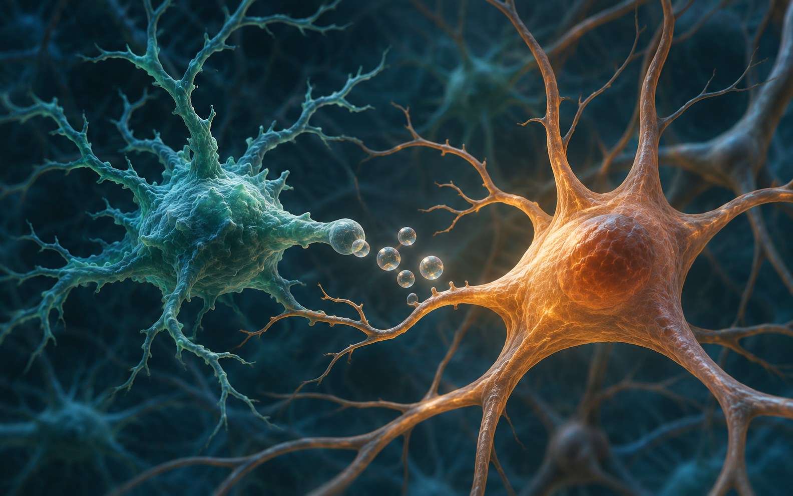
Brain cells send out tiny packets that can carry molecules to other cells. A study in Nature Aging asks whether some of those packets can carry damage as well. In donated brain tissue from people with Alzheimer’s disease, researchers found more of a particular kind of packet and higher levels of an enzyme it carries. In a separate set of mouse experiments, turning down that enzyme eased some Alzheimer’s-like changes.
Evidence level Early Research. The human tissue comparisons were observational. The interventions were tested in mice and laboratory cells.
What travels in a mitovesicle?
Extracellular vesicles are tiny, membrane-wrapped packages that cells release into their surroundings. Mitovesicles are a subset that come from mitochondria, the structures that help cells turn fuel into usable energy. A mitovesicle carries some of the mitochondrion’s contents. When another cell takes up the package, its cargo can affect how that cell works.
Yun Li and colleagues separated vesicles from brain tissue and used microscopy and marker proteins to distinguish mitovesicles from other kinds of vesicles. The team then compared the proteins in mitovesicles from 5×FAD mice, which develop amyloid plaques early, with those from ordinary mice. A protein survey using mass spectrometry flagged an enzyme called gamma-glutamyltransferase 1, or GGT1. Antibody-based tests confirmed higher levels of the enzyme in the mitovesicle fraction from 5×FAD mice. The other vesicle fractions showed no clear difference in GGT1.
Cell experiments pointed to microglia, the brain’s resident immune cells, as a source. After exposure to clumps of amyloid beta, cultured microglia released vesicles with more GGT1. The team saw no comparable increase in vesicles from the neuron or astrocyte cell lines it tested. GGT1 breaks down glutathione, a molecule cells use to manage oxidative stress. That gave the researchers a way to investigate how the cargo might affect recipient cells.
What the human tissue showed
The researchers recovered more mitovesicles from donated Alzheimer’s brain tissue than from tissue from cognitively normal donors. The paper’s figure caption reports four samples for the particle-count comparison. A separate, small comparison found more GGT1 in the isolated mitovesicles from Alzheimer’s tissue. Staining also showed more of the enzyme in microglia in the hippocampus, a brain region important for memory.
These comparisons capture what was present in tissue at the time it was collected. They leave open whether the vesicles helped drive the disease, appeared in response to it, or did some of both.
What changed when the team suppressed GGT1 in mice
When the researchers injected mitovesicles from 5×FAD brains into ordinary mouse brains, the recipients performed worse on learning and memory tasks and showed more inflammation and changes at synapses, the connections between neurons. The vesicles also worsened disease-related changes when injected into mice that already carried the 5×FAD mutations.
The team then switched off the mouse version of GGT1 in microglia in the 5×FAD model. Compared with 5×FAD controls, those mice found a hidden platform faster in a water maze and explored a new arm of a different maze more. The researchers also found less amyloid buildup and lower measures of inflammation. In a separate experiment, the GGT-blocking compound acivicin also eased several disease-related changes. Acivicin acts on targets beyond GGT1, so the microglial gene experiment helps narrow down which enzyme mattered.
What it would take to test a treatment
The experiments give researchers a candidate mechanism to test. The 5×FAD model captures some features of Alzheimer’s, especially amyloid buildup, while the human tissue study cannot show how much this pathway contributes to disease progression in people. Other vesicle cargo could also be involved. The study leaves open whether GGT1 can be safely and selectively blocked in the human brain and whether doing so would improve symptoms or slow decline.
Study Li and colleagues, Nature Aging, November 10, 2026 (DOI 10.1038/s43587-026-01263-3).