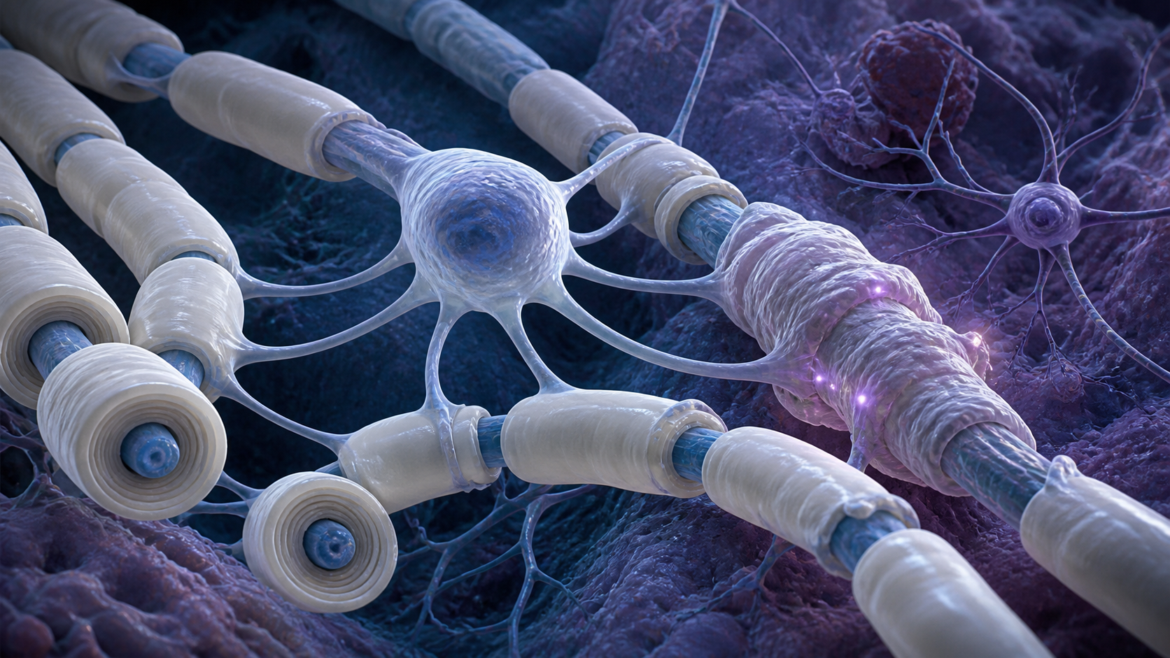
Research on cognitive aging often centers on neurons, the cells that carry electrical signals through the brain. A new study suggests that oligodendrocytes, the cells that produce insulating myelin around nerve fibres, may also be involved. Human brain tissue linked cognitive decline with unusual myelin and oligodendrocyte patterns, while mouse experiments identified reduced NRF2 stress-response activity as one possible mechanism.
Evidence at a glance
- Study type: Human longitudinal observation and postmortem brain analysis, followed by mechanistic mouse experiments.
- Human cohort: Lothian Birth Cohort 1936, with 866 participants followed cognitively from about age 70 to 82.
- Detailed tissue subsets: Small postmortem samples, including 21 brains for one myelin analysis and 15 for electron microscopy.
- Evidence level: Human observational evidence supported by animal experiments.
- Main caution: The study does not establish a treatment for cognitive decline in people.
What oligodendrocytes and myelin do
Oligodendrocytes make myelin, a fatty insulating material wrapped around many nerve fibres. Myelin helps electrical signals travel quickly and reliably. Healthy cognition therefore depends not only on neurons but also on the cells and structures that support communication between them.
Aging is often associated with loss or damage. The new findings were more complicated. People with worse cognitive trajectories had more oligodendrocytes in the examined white matter, smaller myelinated axons and unusually thick myelin. More cells and thicker insulation were not necessarily signs of better function.
How the human study was structured
Participants had taken an intelligence test at age 11 and later completed repeated cognitive assessments roughly every three years between ages 70 and 82. Most showed some decline, but the pace varied. After death, donated brain tissue allowed researchers to compare cognitive trajectories with the condition of white matter.
The longitudinal testing is a major strength because it measures change over time instead of relying on a single late-life score. The detailed tissue analyses, however, involved small subsets. That limits precision and makes independent replication important.
NRF2 links stress response to myelin biology
The researchers found lower activity in an NRF2-related program within oligodendrocytes. NRF2 is a protein that helps cells switch on defenses against oxidative and other forms of cellular stress. When this response is weaker, cells may be less able to maintain normal function under age-related strain.
In aged mice, removing NRF2 specifically from oligodendrocytes produced changes resembling the human tissue findings. The manipulation altered oligodendrocyte and myelin features and reduced the cognitive improvement otherwise seen in part of the experiment. This gives the NRF2 connection mechanistic support, but the evidence comes from animals.
Why the result is not a human treatment
The human component shows a relationship among cognitive decline, oligodendrocytes, myelin structure and NRF2 activity. It cannot determine which change came first. Extra oligodendrocytes might represent a failed repair response, a consequence of other brain pathology or part of the process contributing to decline.
The mouse work tests causality more directly, but mouse white matter, lifespan and cognitive tasks are not identical to those of people. It would be unsafe to conclude that reducing oligodendrocytes, thinning myelin or activating NRF2 will prevent human cognitive decline. A useful intervention would need to restore healthy tissue function without disrupting the essential insulation that myelin provides.
Important limitations
The cohort was drawn largely from white European participants in Scotland. The people who donated brain tissue may also differ from the wider population. Postmortem studies capture one biological endpoint and cannot show how every tissue change developed during life.
Future research should reproduce the findings in larger and more diverse brain banks, connect tissue measures with imaging obtained during life and test whether the identified cell states appear in other forms of cognitive aging.
The Lifespan Brief assessment
The study broadens the biology of cognitive decline beyond neurons. It supports the idea that the brain’s support cells and wiring insulation can become maladaptive with age. The combined human and mouse evidence makes NRF2-related oligodendrocyte dysfunction a credible research target, but not a proven therapy.
Primary source
Oligodendrocyte dysfunction in human age-related cognitive decline, Nature Medicine, published August 25, 2026.