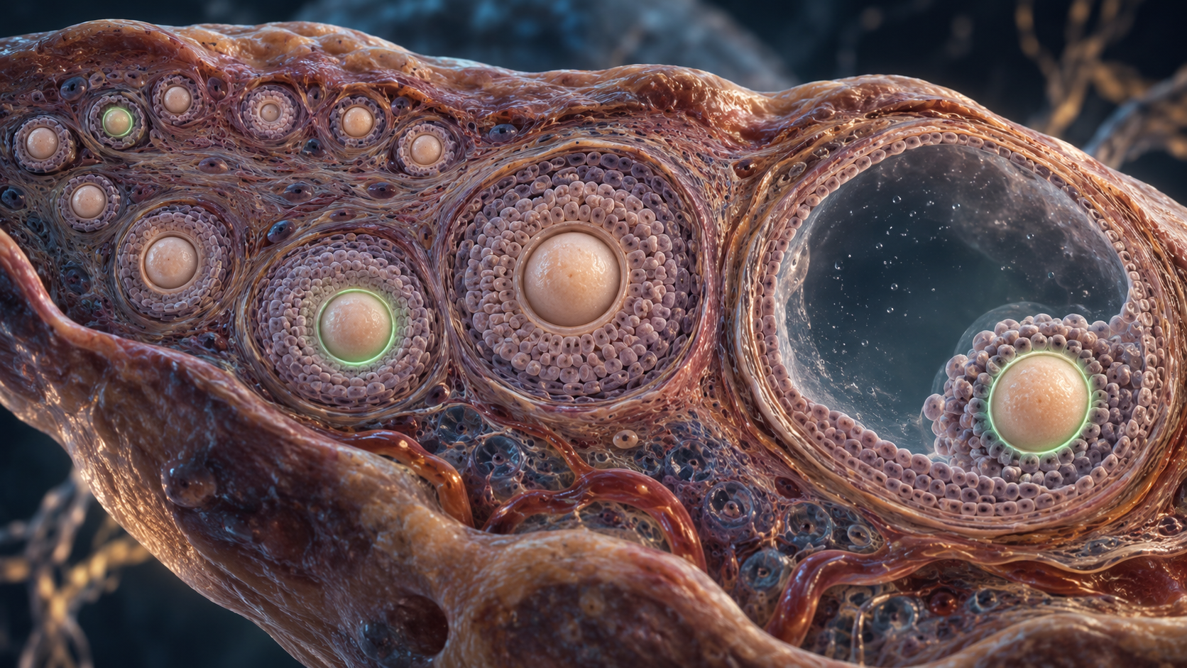
An egg cell that has begun developing does not necessarily rush toward ovulation. New mouse research finds that some activated oocytes can remain in growing follicles for much of the animal’s reproductive lifespan, while retaining the ability to produce offspring.
The finding challenges a simple view of ovarian aging as a countdown controlled only by the dormant egg supply. It suggests that the pace of development after activation also helps regulate how that supply is used.
The paper behind the finding
Jing Liang, Xuebing Yang and colleagues, led by corresponding author Hua Zhang, published the open-access study in Nature Aging on October 8, 2026. The full paper is publicly accessible. Its experiments examine mouse reproductive biology, not a treatment to extend fertility in women.
What is an ovarian follicle?
An oocyte is an immature egg cell. A follicle consists of that cell and the surrounding support cells that help it develop. Small primordial follicles hold dormant oocytes. After activation, follicles pass through primary and secondary stages before forming the fluid-filled antral structures associated with later maturation.
The ovarian reserve is finite. Many follicles are lost rather than ovulated, and both egg quantity and quality matter for reproductive aging. Understanding when follicles grow, pause or disappear is therefore more useful than treating the reserve as a single number.
Following a labeled group over time
The researchers used an inducible genetic tracing system based on Zp3, a gene active in growing oocytes. A brief tamoxifen exposure switched a fluorescent reporter from red to green in activated oocytes, while dormant oocytes remained red. That let the team identify the labeled group months later.
They labeled oocytes at postnatal day 35 and examined ovaries at successive time points across 12 months. This was cohort tracing with tissue collection, not continuous filming of every individual follicle. Key time-course experiments used five mice per time point.
About 48% of the initially labeled oocytes remained detectable after two months, about 15% after six months, and roughly 4% after 12 months. Those percentages refer to the initially labeled group, not to all eggs in the ovary.
The secondary follicle as a buffer
Labeled oocytes disappeared from primary follicles within about a month. Secondary follicles behaved differently: some developed quickly, while others retained labeled oocytes for many months. Around 80% of the persistent labeled oocytes in the secondary and antral compartments were in secondary follicles.
The authors infer that secondary follicles can persist for at least 11 months in this mouse model. They describe this compartment as a “secondary ovarian reserve,” a developmental buffer that spreads oocyte use across time. It is an existing pool of developing eggs, not evidence that the adult ovary makes replacement eggs.
The long-lived cells were not merely fluorescent remnants. Labeled oocytes recovered months later could be fertilized and produce viable offspring. Natural mating also produced labeled offspring at six and nine months after labeling.
How growth rates may be controlled
Single-oocyte protein measurements compared six long-lived labeled oocytes with six unlabeled oocytes. The long-lived group showed lower biosynthetic and metabolic activity, alongside reduced proliferation of surrounding granulosa support cells. The small protein-analysis sample makes broader replication valuable.
The team also investigated FURIN, an enzyme that processes signaling proteins. Genetic experiments linked it to oocyte-secreted factors, including GDF9, and communication with surrounding follicle cells. Removing FURIN reduced follicle-cell proliferation and caused secondary follicle arrest. That result identifies a control mechanism, but arrest is not the same as preserving useful fertility.
Oocytes labeled in older mice disappeared within about three months. In young mice with one ovary surgically removed, the labeled group was depleted within about seven months. These results support flexible utilization, although surgery changes more than reserve size alone.
What this changes, and what remains unknown
The study expands attention from dormant follicles to the timing of development after activation. It differs from the earlier whole-ovary mapping study we covered, which measured the distribution of follicle states. Here, labeling reveals how long an activated group can persist.
Whether human follicles have comparable long-term dynamics remains unverified. The authors’ suggestions about years or decades of development in people are hypotheses extrapolated from mice. Controlled laboratory conditions also differ from human exposures.
The Lifespan Brief verdict
This is a meaningful revision of the mouse follicle-development timeline. It provides a new framework for studying reproductive aging, but demonstrates no established method for extending human fertility, delaying menopause or improving human lifespan.
Feature image: AI-generated conceptual illustration of ovarian follicles and fluorescent labeling, not experimental microscopy.