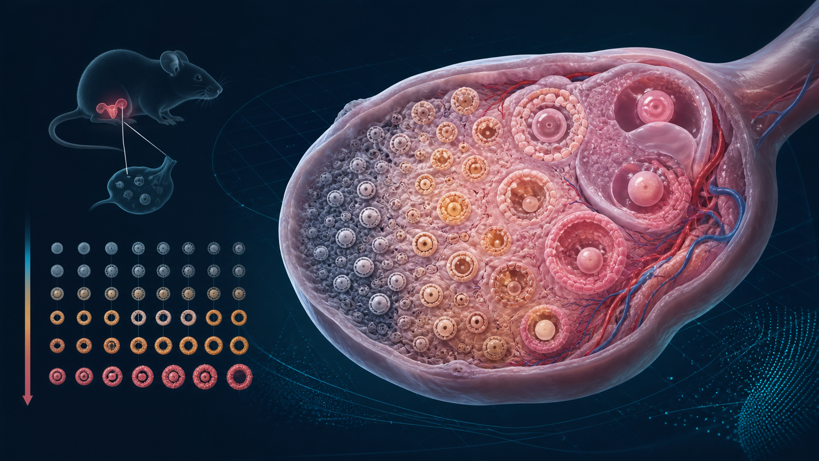
A three-dimensional map of more than 85,000 oocytes across the mouse reproductive lifespan has revealed an unexpected pattern. Even as the ovarian reserve fell by more than tenfold, about 14% of the remaining oocytes stayed in a transitional state between dormancy and growth.
The countdown built into reproductive aging
An oocyte is an immature egg cell. Oocytes sit inside follicles, structures made up of the oocyte and surrounding support cells. Together, the dormant and developing follicles in the ovaries make up the ovarian reserve.
Unlike most cell populations, this reserve is largely established before birth and is not meaningfully renewed during adult life. In humans, the ovaries contain roughly one to two million oocytes at birth, about 400,000 by puberty and around 1,000 by menopause. Only a small fraction are ever ovulated. Most are lost through normal biological processes.
This built-in depletion makes reproductive aging different from aging in many other organs. Fertility can decline decades before other systems fail, and the ovaries also produce hormones that influence bone, cardiovascular and metabolic health. That is why ovarian aging has become an increasingly important part of geroscience, the study of how aging biology drives disease and loss of function.
Mapping whole ovaries in three dimensions
Traditional follicle counts usually rely on thin two-dimensional tissue slices. Those sections can provide excellent detail, but they sacrifice the geometry of the intact organ and require estimates to reconstruct the total reserve.
For a study published in Nature Aging on August 12, 2026, researchers combined light-sheet microscopy with artificial intelligence-based image segmentation. Light-sheet microscopy illuminates a thin plane inside a cleared tissue sample, allowing the whole organ to be imaged layer by layer. The software then identified and measured oocytes throughout each ovary.
The team analyzed more than 100 ovaries from 56 inbred C57BL/6J mice ranging from five to 60 weeks of age. In total, the pipeline segmented more than 85,000 oocytes. The researchers classified them by size and by the shape of the granulosa cells surrounding them, distinguishing dormant primordial follicles, transitional follicles and growing follicles.
The surprising 14% pattern
As expected, the total number of oocytes declined sharply with age. The balance of developmental stages also shifted. Primordial oocytes fell from about 70% to about 55% of the reserve, while growing oocytes increased from about 15% to about 30%.
The transitional group behaved differently. Across every age examined, transitioning oocytes remained at roughly 14% of the total reserve. This stability persisted even while the overall number of oocytes dropped by more than an order of magnitude.
A transitional follicle has started moving away from dormancy but has not yet reached the fully growing primary stage. Maintaining a similar proportion of these follicles suggests that recruitment is not simply a fixed number of oocytes switching on each week. Instead, the ovary may regulate activation in relation to the size of the remaining reserve.
Is the ovary sensing its remaining reserve?
The authors describe the pattern as evidence for an organ-scale regulatory mechanism. Their spatial analysis also found that regions with a higher density of primordial oocytes tended to contain more recently activated oocytes. That observation argues against a simple model in which crowded follicles inhibit each other from activating.
A mathematical model fit to the data estimated that about 2.5% of primordial oocytes entered the next developmental stage each week. It also predicted that most oocyte loss occurred later, at the antral stage, rather than during the primordial, primary or secondary stages.
Models are useful for testing whether a set of biological rules can reproduce observed data. They are not direct measurements of every transition, and the authors noted that their model assumes growth and death rates remain broadly similar across aging ovaries. Future experiments will need to identify the signals that might allow the tissue to coordinate recruitment.
Why genetically identical mice differed so much
The researchers also found large differences in total oocyte numbers among mice of the same age, even though the animals were genetically identical and raised under similar conditions. The variation was already visible before sexual maturity.
That timing suggests the starting reserve may be shaped by developmental events. Small differences in the number of early germ cells, their migration into the developing gonad or the loss of oocytes around birth could establish different baselines. The study did not determine which factor was responsible.
This variability matters because it cautions against treating ovarian reserve as a simple genetic countdown. Developmental history, local tissue conditions and chance may all influence the number of follicles present at a given age.
The connection to broader longevity research
Researchers are increasingly asking whether preserving ovarian function could affect health beyond fertility. Menopause changes hormone exposure throughout the body, and earlier menopause is associated with several later-life health risks. Those associations do not prove that extending ovarian function would extend lifespan or prevent disease.
The immediate value of this study is more basic. It provides a quantitative framework for asking how a whole organ manages a finite cell reserve over time. It also shows why spatial organization matters. Counting cells alone can miss relationships among follicle density, location and developmental state.
The imaging and segmentation approach was also applied to small pieces of ovarian cortex from two 25-year-old human donors. That demonstrates technical feasibility in human tissue, but it does not reproduce the mouse lifespan analysis in people. The stable 14% pattern, the age comparisons and the regulatory interpretation all come primarily from mice.
Why this does not mean menopause can be delayed
- The study did not manipulate the transitional oocyte population.
- It did not show that changing the 14% proportion extends reproductive lifespan.
- It did not demonstrate a way to delay, prevent or reverse menopause in humans.
- It did not show that preserving ovarian function increases overall lifespan.
- The large reserve differences among genetically identical mice do not identify their cause.
Even if researchers identify the signals that regulate follicle activation, intervening safely would be difficult. Keeping more follicles dormant might preserve quantity while affecting oocyte quality, ovulation or hormone production. Reproductive aging is not controlled by a single counter.
The Lifespan Brief verdict
This is a meaningful advance in understanding ovarian aging. Whole-organ imaging, a large age-spanning dataset and mathematical modeling revealed a stable transitional fraction that traditional tissue sections could easily miss. The result supports the idea that follicle recruitment is coordinated at the tissue level. But the central evidence comes from mice, and the study does not demonstrate a human menopause or longevity intervention.