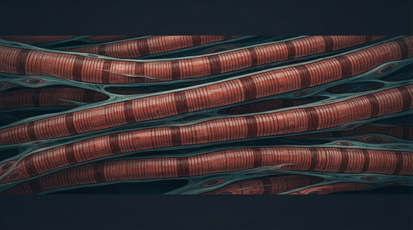
Female mice lived about three months longer at the median when a gene called Terf2 was deleted from their skeletal muscle fibres early in life. At 15 months, their fibres also showed less age-related shrinkage, and their muscles produced more force early in a fatigue test. Male mice showed no comparable survival benefit.
The study, published on November 12 in Philosophical Transactions of the Royal Society B, started with an observation that seems counterintuitive. TRF2, the protein made by Terf2, helps protect the ends of chromosomes, yet its levels naturally decline in skeletal muscle from early adulthood. The researchers wanted to see what happens to muscle over time when that protein is missing from the fibres.
What changed in the muscles
Muscle fibres generally get smaller with age as protein turnover, repair and the nerve signals that drive contraction change. Losing fibre size can contribute to weakness, though strength also depends on how well the nervous system recruits the muscle that remains.
The team used a mouse model in which Terf2 was switched off in skeletal muscle fibres early in life. They compared the mice with controls, examined two lower-leg muscles under a microscope and measured the force produced by the muscles that push the foot downward. They followed separate groups for survival.
At 15 months, the measured muscle fibres in the modified mice had largely retained their cross-sectional size, while fibres in the controls had shrunk. Modified female mice had a modest advantage in initial strength and produced more force early in repeated contractions. By the end of the fatigue test, a control group held up better. The result points to an advantage in early force production, with a trade-off in sustained performance under this test.
How much longer the mice lived
Median survival was 11% longer in the modified females, a gain of about three months. The survival comparison included 23 modified females and 27 female controls. Both male groups had a median lifespan of around 25 months. The survival result measures the midpoint of the groups’ lifespans and does not establish that changes in muscle performance caused the difference.
Why the loss of a protective protein might help
Telomeres are stretches of DNA and associated proteins that cap chromosome ends. TRF2 helps shield those ends from being treated as broken DNA. In the modified mice, the researchers found no significant rise in the telomere damage markers they measured in skeletal muscle at 15 months.
Gene activity in female muscle pointed to changes in pathways involved in how mitochondria produce energy. The researchers proposed that mild oxidative stress may trigger protective responses, an idea known as hormesis. Earlier work from the group identified FOXO3a as a possible backup for telomere protection in muscle fibres. The new study did not directly show that mitochondrial stress or this backup mechanism accounted for the longer survival.
What the study leaves open
Some muscle measurements involved only a handful of mice per group, and the study assessed muscle function at 15 months. The gene deletion began early in life, giving the mice time to adapt. Whether reducing TRF2 in adult muscle would reproduce the effects, and whether any such approach could be safe or effective in humans, remain open questions.
Original research · Gilson and colleagues in Philosophical Transactions of the Royal Society B (open access).
Related reading · What Telomere Length Can Tell Us About Aging in Midlife