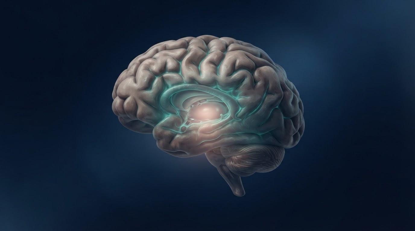
The hypothalamus helps keep the body on schedule. It coordinates sleep and waking, helps regulate hunger and energy use, and links the brain to hormonal responses to stress. A study in Nature maps how different parts of this small structure change with age and identifies shifts in gene activity in some of the cells that help set the body’s daily rhythms in people with early Alzheimer’s disease.
One region, many jobs
The hypothalamus sits near the base of the brain. It contains clusters of cells called nuclei, each with a somewhat different job. The suprachiasmatic nucleus serves as the body’s central clock, helping synchronize daily rhythms with the light-dark cycle. The arcuate nucleus helps regulate appetite and energy balance. Looking at the size of the hypothalamus as a whole can obscure changes within these much smaller structures.
The researchers built an atlas called HypoAD using high-resolution MRI scans from 458 people and gene-expression profiles covering 614,403 cell nuclei. MRI showed how the size of hypothalamic structures varied across people. Single-nucleus RNA sequencing measured which genes were active in different types of cells from donated brain tissue. Here, the word nucleus has two meanings. On the scans it refers to a cluster of brain cells. In the sequencing work it refers to the compartment inside an individual cell that houses its DNA.
Ageing changed the shape of the map
Some hypothalamic structures became smaller with age, including parts of the lateral hypothalamus. Others, including the arcuate and suprachiasmatic nuclei, showed signs of being larger in older adults after the researchers accounted for measures of amyloid buildup and cognitive impairment. Differences associated with cognitive impairment and amyloid were generally smaller or harder to detect on MRI than differences associated with age.
Volume alone gives limited information about what is happening inside a structure. A slightly larger region could reflect changes in cell size or in supporting tissue, among other possibilities. The team also flagged the suprachiasmatic nucleus as especially difficult to measure reliably on MRI. Its size estimates warrant particular caution.
The signals that stood out in Alzheimer’s
For the post-mortem Alzheimer’s comparison, the team profiled tissue from 14 donors, seven with Alzheimer’s and seven controls. One donor with familial Alzheimer’s was included as a distinct example and left out of the formal comparisons. The molecular data showed changes in some inhibitory neurons, which help regulate the activity of other neurons. The analysis pointed to populations involved in circadian timing and sleep, including cells the researchers mapped to the suprachiasmatic and dorsomedial regions of the hypothalamus.
Microglia, the brain’s resident immune cells, also showed shifts in the activity of genes involved in inflammation. In the clock-related neurons, some changes involved signalling pathways that help coordinate daily rhythms. These findings give researchers candidate pathways to investigate as they work to understand why sleep and daily activity can become disrupted in Alzheimer’s. The study did not track those symptoms alongside the cellular changes in the same individuals.
The MRI and molecular data came from different groups of people. The study therefore could not tie a change seen on a scan to a particular cell change in the same person. It also captured snapshots across participants and donated tissue. Establishing when these changes emerge will require studies that follow people over time and, where possible, pair more precise imaging with measurements that can validate what is happening in the tissue.
The atlas makes it possible to ask more precise questions than whether the hypothalamus as a whole is affected. A future study could examine whether changes in the central clock track the fragmentation of sleep and waking in the same people. Another could test whether the microglial signals appear around the same time as changes in sleep-related neurons. The order matters because an early change and a late response to disease point to different biological explanations.
Primary source: Nature original study, November 18, 2026.
The Takeaway
HypoAD gives researchers a reason to look more closely at the brain’s timekeeping circuits in Alzheimer’s. The anatomical work shows that ageing affects parts of the hypothalamus differently, while the tissue analysis points to changes in specific clock-related neurons and immune cells in early disease. The next question is whether those cellular changes precede or follow the sleep and rhythm disruptions people experience, and whether the two can be connected in the same individuals.