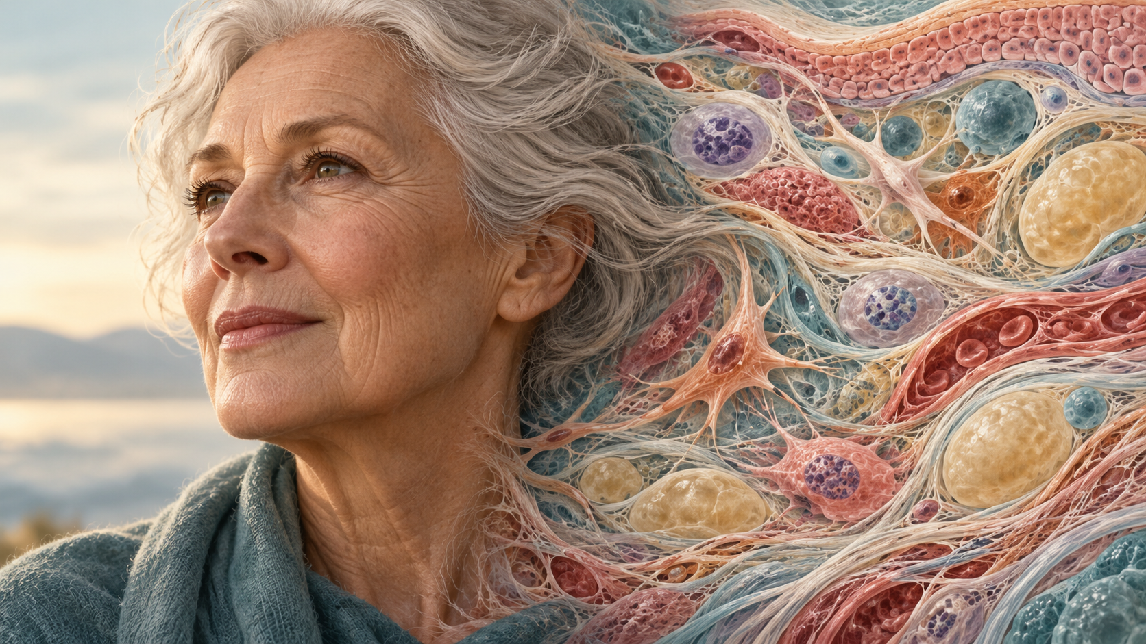
The short answer: Cells are replaced at very different rates, and replacement does not erase all the changes that develop with age. Some cells remain for decades. New cells can inherit DNA changes from the cells that produced them, and their function depends partly on the condition of the surrounding tissue.
If the body can replace worn-out cells, why do we get wrinkles, lose strength or become more vulnerable to disease? The question makes sense when we hear how much renewal happens every day.
To understand how renewal and aging happen together, it helps to look at which cells are replaced, where their replacements come from and what happens to the tissue around them.
The seven-year replacement myth
You may have heard that every cell in your body is replaced every seven years. There is no such body-wide schedule.
A 2021 analysis by Ron Sender and Ron Milo estimated daily turnover at roughly 330 billion cells in a reference healthy young adult man. Most of those cells were blood cells, with gut-lining cells making up much of the rest. The researchers combined measurements from earlier studies, so the estimate should not be treated as an exact daily count for everyone.
Many gut-lining cells last only a few days, while other cell populations persist for years. Rapidly renewing populations account for much of the daily total.
Over time, the same short-lived cell populations are replaced repeatedly. Adding up those replacements would overstate how much of the rest of the body had been renewed.
Some of your cells are nearly as old as you
Researchers have estimated the age of cells using carbon-14 left in the atmosphere by Cold War nuclear tests. Carbon entered the food chain and became incorporated into newly made DNA, helping scientists estimate when cells formed.
In a human study published in 2006, neurons in the cerebral neocortex, the brain’s large outer region, were largely as old as the people they belonged to. The researchers found no detectable substantial replacement in adulthood. The study examined that region specifically; other brain regions and cell types can have different renewal patterns.
A 2009 study of human heart muscle likewise found renewal, but at a slow rate that declined with age. Its model estimated annual replacement at about 1% at age 20 and 0.3% at age 75. These estimates are inconsistent with complete replacement of heart muscle cells every seven years.
Long-lived cells still maintain themselves. Replacing proteins or other components inside a cell is different from replacing the entire cell. A cell’s age is therefore not a simple measure of how recently everything inside it was made.
New cells can inherit DNA changes
Where replacement does happen, the next generation usually comes from cells already living in your body. Many tissues rely on stem cells, which can maintain their own population while producing specialized descendants.
Stem cells themselves acquire changes over a person’s lifetime.
In a 2016 study, scientists grew small tissue cultures from individual human stem cells taken from the small intestine, colon and liver. Across 19 donors aged 3 to 87, they found that DNA mutations accumulated with age.
A mutation is a change in the DNA sequence. If a lasting change is present in a stem cell, it can be passed on when that cell divides. The daughter cell may therefore carry a mutation acquired earlier in the life of its parent cell.
Many mutations have little or no effect on cell function. This study also cannot establish how much mutation accumulation contributes to aging overall. It does, however, show that genetic changes can persist in tissues that continue producing new cells.
Stem cells change with age
Stem cells must do more than divide. They need to remain available, respond at the right time and produce the right kinds of cells. Their surroundings help regulate that balance.
A 2012 mouse study illustrates the problem. Older muscle fibers produced more of a growth signal called FGF2. This pushed some nearby muscle stem cells out of their resting state and contributed to depletion of the stem-cell pool.
A resting stem cell remains available for future repair. In these experiments, prolonged activation reduced the pool of cells able to help when muscle was injured.
The findings come from mice and would need further investigation before they could guide treatment in people. Within this model, stem-cell preservation depended on regulating when cells entered division.
Surrounding tissue influences cell behavior

Cells attach to supporting material, respond to chemical messages and interact with neighboring cells. The extracellular matrix is the network of proteins and other material around them, including collagen.
In research using human skin and laboratory tissue models, fragmented collagen altered how fibroblasts, cells that maintain connective tissue, stretched and functioned. Cells in damaged collagen environments showed increased oxidative stress and production of a collagen-degrading enzyme.
These experiments suggest that changes in the surrounding structure can impair cell function. Replacing cells alone may leave those tissue changes in place.
Some biological material also lasts exceptionally long. Radiocarbon dating of human eye lenses found that proteins in the lens core were formed largely around birth and retained throughout life. The lens grows, but its center is not routinely cleared out and rebuilt.
The lens provides another example of how much renewal varies within the body, down to particular regions of an organ.
Why not just make everything divide faster?
Cell division needs controls that prevent abnormal growth and help tissues retain their normal structure.
One protective response to cellular stress is senescence: cells stop dividing and enter a changed state. This can help prevent damaged cells from continuing to multiply. But some senescent cells also release signals that affect surrounding tissue.
In experiments reported in 2008, senescent human cells released inflammatory and other signaling molecules. Their secretions changed the behavior of nearby cells in culture. Much of this work concerned cancer-related mechanisms, so it should not be read as proof of a treatment for ordinary human aging.
Senescent cells can therefore influence tissue even after they stop dividing. Understanding those effects requires examining both the cells being replaced and the signals produced by cells that remain.
The Lifespan Brief answer
Cell replacement helps tissues continue functioning, but several sources of age-related change can persist through that renewal.
Some cells and molecules remain for much of a lifetime. Replacement cells can inherit changes from their predecessors. Stem-cell systems can lose capacity or receive inappropriate instructions. Supporting tissue can change, while stressed cells can alter the signals around them.
The contribution of each process differs among tissues. Researchers are still investigating how they interact, which changes drive further damage and which arise as responses to it.
The body continues producing cells throughout life while also retaining long-lived cells, accumulated genetic changes and altered tissue structures. Those processes can occur together, allowing renewal to sustain an organ without restoring its earlier condition.
For aging research, this means that the number of new cells is only one measure to examine. Their function, the condition of the cells producing them and their ability to integrate into tissue also need to be assessed.
Sources
Sender R, Milo R. The distribution of cellular turnover in the human body. Nature Medicine (2021). Original publication · Freely accessible full paper.
Bhardwaj RD et al. Neocortical neurogenesis in humans is restricted to development. PNAS (2006). Human tissue research.
Bergmann O et al. Evidence for cardiomyocyte renewal in humans. Science (2009). Human tissue research and mathematical modeling.
Blokzijl F et al. Tissue-specific mutation accumulation in human adult stem cells during life. Nature (2016). Human stem cells expanded as organoid cultures.
Chakkalakal JV et al. The aged niche disrupts muscle stem cell quiescence. Nature (2012). Mouse experiments.
Fisher GJ et al. Collagen fragmentation promotes oxidative stress and elevates matrix metalloproteinase-1 in fibroblasts in aged human skin. American Journal of Pathology (2009). Human skin and laboratory tissue models.
Lynnerup N et al. Radiocarbon dating of the human eye lens crystallines reveal proteins without carbon turnover throughout life. PLOS ONE (2008). Human tissue research.
Coppé J-P et al. Senescence-associated secretory phenotypes reveal cell-nonautonomous functions of oncogenic RAS and the p53 tumor suppressor. PLOS Biology (2008). Primarily cultured human cells, with patient-tissue observations.
Images are original AI-generated editorial illustrations for The Lifespan Brief. They are conceptual, not microscopy or study data. This article is for general information and is not personal medical advice.